
Lesson plan "type of flatworms". Comparisons of coelenterates and flatworms. Body symmetry. Presence of layers of cells. Body Shape
Examples of worksheets for a general lesson on the topic “Worms”
Worksheet
Crossword.
Horizontally:
Exercise 1. Color the drawing of the planarian worm's organ system. The organs of the nervous system are yellow, the digestive organs are green, and the excretory organs are blue. Name the organs and their functions.
Task 2. Compare the structure of animals of the Coelenterate type and the Flatworm type according to the plan. Identify progressive features in development flatworms. Draw a conclusion about the place of flatworms in historical development animal world.
Plan for comparison of coelenterates and flatworms.
1. Body symmetry.
2. Presence of layers of cells.
3. Body shape.
4. Presence of tissues, organs and organ systems.
5. Removing undigested food debris.
6. Ability to regenerate.
7. Breathing.
Task 3. Creative task. Bovine and pork tapeworms are found on almost all continents and large islands, i.e. they are cosmopolitan animals. What is the reason for their wide geographical distribution?
Task 4.
Task 5. “Who is who”
Worksheet
Crossword
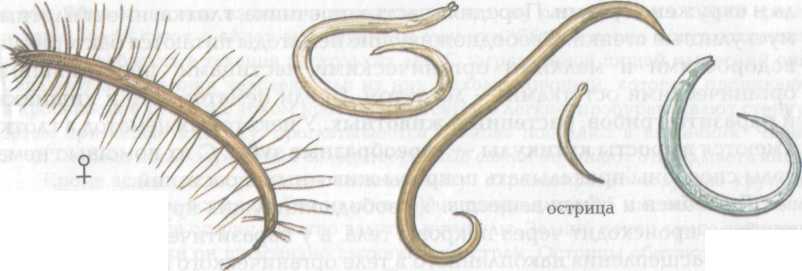
Horizontally: 1. An animal whose body contains both female and male reproductive organs. 8. Loose connective tissue that fills the spaces between organs in flatworms. 9. Inner layer cells. 10. Body cavity filled with fluid in roundworms. 11. Name of the class in the phylum Roundworms.
Exercise 1. Color the drawing of the organ system of the roundworm worm. The organs of the nervous system are yellow, the digestive organs are green, and the excretory organs are blue. Name the organs and their functions.
Task 2. Compare flatworms with roundworms according to plan. Identify progressive features in the structure of roundworms. Draw a conclusion about the place of roundworms in the process of evolution.
Plan for comparison of round and flatworms.
1. Body symmetry.
2. Layers of cells that form the body.
3. Body shape.
4. Body cavity.
5. Skin-muscle bag.
6. The structure of the digestive system.
Task 3. Creative task. There are roundworms whose microscopic larvae cause elephantiasis. Adult worms clog the lymphatic vessels, their walls thicken, and lymph stagnation occurs. In tropical and subtropical areas where malaria is actively combated, this disease practically does not occur. How can this be explained?
Task 4.
Formulate measures to prevent helminth infection.
Task 5. “Who is who”
Here are images of worms. Name what kind of worms they are, what type and class they belong to. Tell us what role they play in nature or human life.
Worksheet
Crossword
Horizontally: 1. An animal whose body contains both female and male reproductive organs. 8. Loose connective tissue that fills the spaces between organs in flatworms. 9. Inner layer of cells. 10. Body cavity filled with fluid in roundworms. 11. Name of the class in the phylum Roundworms.
Exercise 1. Color the drawing of the earthworm's organ system. The organs of the nervous system are yellow, the digestive organs are green, the excretory organs are blue, and the circulatory organs are red. Name the organs and their functions.
Task 2.
Identify common and progressive features in the structure annelids compared to other worms on the plan. Draw a conclusion about the place of annelids in the evolutionary tree of animals.
A plan for comparing annelids to other worms.
1. Body symmetry.
2. Body shape.
3. Dividing the body into sections.
4. Organs of locomotion.
5. Body cavity.
6. Circulatory system.
7. Digestive system.
8. Excretory organs of flatworms and annelids.
9. Ability to regenerate
Task 3.Creative task. At the end of the 19th century, one doctor, in an experiment carried out on himself, found out that larvae and subsequently adult worms do not develop from eggs just laid by a female roundworm and swallowed by a person. How can we explain the results obtained in the experiment? How do roundworms stay in the host’s body, since they don’t have attachment organs like flatworms?
Task 4.
Formulate measures to prevent helminth infection.
Task 5. “Who is who”
Here are images of worms. Name what kind of worms they are, what type and class they belong to. Tell us what role they play in nature or human life.
Test work
Test work
Test work
Crosswords
6-8 grades
Self-analysis of the lesson on the topic “Worms”
“We have learned to swim in the water like fish, fly in the sky like birds, all that remains is to learn to live on Earth like people!”
Bernard Show.
Table 2.
Comparative characteristics sponges and coelenterates
Topic 7. Type Ctenophora
Theoretical part
Ctenophores are marine, predominantly swimming animals, less often crawling or sessile. In total, about 120 species of ctenophores are known. In 1888 Ctenophores were identified as a separate phylum.
Ctenophores belong to the Radiata section and have some common features: radial symmetry, two-layer structure and the presence of an intestinal cavity. A specific feature of ctenophores is the presence of special organs of movement - paddle plates located in meridional rows. Each pectal plate consists of adherent large cilia of the epithelium. Ctenophores lack stinging cells, but have special adhesive cells concentrated on the hunting tentacles. Ctenophores have a special aboral organ that regulates movement.
The development of ctenophores is direct, without passing through the larval phase. During the process of embryogenesis in ctenophores, in addition to the ectoderm and endoderm, the rudiment of the third germ layer is laid - the mesoderm, from which the muscles of the tentacles and mesoglea cells are formed.
The phylum Ctenophores includes only one class: the class Ctenophores.
Ctenophores inhabit all seas. Among them, the smallest reach 2-3 mm in length, and the largest, like the Venus belt, can be up to 3 m long. Most ctenophores feed on plankton.
The most typical floating forms are oval with tentacles and ribbon-shaped without tentacles. Specialized forms are crawling and sessile ctenophores.
Swimming ctenophores have a mouth on the oral pole of the body, and an aboral organ on the aboral pole.
Ctenophores move almost exclusively due to the flapping of the rowing plates, and the contraction of muscle fibers allows them to change the direction of movement in the water. Muscle cells are most developed in the tentacles, which can strongly stretch and contract. The tentacles have many sticky cells, with the help of which planktonic animals are caught.
The aboral organ functions as an organ of balance and regulates the movement of the rowing plates. The nervous system of ctenophores is diffuse. It is represented by individual nerve cells. Clusters of nerve cells are observed under the rows of paddle plates, near the mouth and under the aboral organ.
Ctenophores have a highly developed mesoglea. Many ctenophores are characterized by a glow in the dark, which helps individuals of the same species find each other in marine spaces.
The gastrovascular system of ctenophores is complex and consists of an ectodermal pharynx and an endodermal stomach with canals extending from it. The mouth of ctenophores is always located at the oral pole and leads into the pharynx. The pharynx goes into the stomach. Channels extend from the stomach. One canal is directed toward the aboral pole and at the end branches into four short branches, two of which are blindly closed, and two open with pores outward. The two blind canals of the gastrovascular system are directed from the stomach to the oral pole and are located on the sides of the pharynx. Two more canals extend from the stomach in the equatorial plane and are divided twice dichotomously. The resulting eight radial channels flow into eight meridional channels, blindly ending at the poles.
Meridional channels lie under the rows of rows and provide the movement of rowing plates with nutrients. Food is captured by the tentacles and transferred to the mouth. Digestion of food occurs in the large pharynx and stomach under the action of digestive enzymes secreted by the glandular cells of the endoderm, then the food is transported through the channels of the gastrovascular system, where it is absorbed by the digestive cells of the endoderm. In addition to the digestive and transport functions, the gastrovascular system performs the functions of an internal cavity where gas exchange occurs, the release of metabolic products occurs, and sexual function occurs.
Ctenophores are hermaphrodites and reproduce only sexually. In the walls of each meridional canal, the male and female gonads are located on the sides. When the gonads mature, germ cells enter the gastrovascular system through tissue ruptures and exit through the mouth. In some species of ctenophores, fertilization of eggs and the subsequent development of zygotes occurs in water, while in others, in the gastrovascular system. Development is direct, without larvae.
Practical part
Questions for self-study
1. What features in the structure of ctenophores made it possible to distinguish them into an independent type?
2. Which germ layer takes part in the formation of mesoglea cells in ctenophores?
3. What is common in the structure of: a) ctenophores and scyphojellyfish; b) ctenophores and coral polyps?
4. The structure of the nervous system of ctenophores.
5. Reproduction of ctenophores.
Topic 8. Type Flatworms (Plathelminthes)
Theoretical part
Flatworms are characterized by the following organizational features.
1. The surface of the body is covered with single-layer ciliated ciliated epithelium. Under the skin there are several layers of muscles: circular, diagonal and longitudinal. Between the dorsal and abdominal walls of the body there are often bundles of dorsoventral muscles that flatten the body. Complex musculature allows for a variety of types of movement.
2. Flatworms do not have a body cavity. All spaces between the internal organs are filled with a special loose tissue - parenchyma.
3. The digestive system consists of two sections: anterior and middle. The intestine is blindly closed, often branched. There is no hindgut or anus. Some primitive forms lack an intestine.
4. The nervous system is of the orthogonal type, which consists of a paired cerebral ganglion and several pairs of nerve cords extending from it, connected to each other by ring cords - commissures. Overall, the nervous system resembles a grid.
5. Sense organs are most developed in free-living species. Many of them have eyes, balance organs - statocysts and numerous sensilla: tactile cells and chemical sense organs.
6. The excretory system of flatworms is represented by individual parenchyma cells, in which excreta accumulate, and protonephridia - branching channels that remove excess fluid from the body with metabolic products dissolved in it. At the inner ends, the excretory tubules end in stellate cells with a “flickering flame.” The protonephridial tubules are connected into one or two excretory canals that open to the outside with excretory pores.
7. Flatworms do not have a circulatory or respiratory system.
8. Flatworms are hermaphrodites, i.e. each individual has male and female gonads. The reproductive ducts are complex. The female reproductive system of most flatworms is characterized by the presence of vitelline glands - glands that produce yolk cells. Their energy material is used by developing eggs. Fertilization is internal.
The phylum Flatworms include nine classes. The most numerous are the class Ciliated worms, the class Flukes, the class Monogenea and the class Tapeworms, or Cestodes.
Practical part
Questions for self-study
1. Which body cavity is called primary?
2. What is the body cavity of flatworms?
3. What is tegument? What worms does it occur in?
4. How do worms without intestines feed?
6. Representatives of what class develop with a change of owners?
7. How do free-living worms breathe?
8. The structure of the nervous system of flatworms?
9. In which worms does asexual reproduction occur? How does it happen?
10. Who is the intermediate and final owner:
a) liver fluke; b) pork tapeworm; c) echinococcal tapeworm;
d) wide tapeworm?
LABORATORY WORK No. 4.
Subject: STRUCTURE OF CILIA WORMS AND FLUKES
Type FLAT WORMS (PLATHELMINTHES)
Class cilia worms
Representative: Mnogoglazka
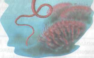
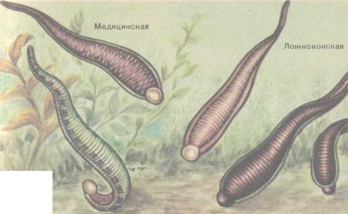
Class DIGENETIC FLUKES
Representatives: Liver fluke, lanceolate fluke.
Goals: to study the structural features of ciliated worms and digenetic flukes, to ensure that their structure corresponds to bilateral symmetry.
Materials and equipment: total microslides of a ciliated worm, microslides of a cross section of a ciliated worm, total preparations of flukes, microscopes, binocular magnifiers.
Progress:
1. Using a magnifying glass, examine and sketch appearance ciliated worm and the structure of the digestive system. Mark the mouth, pharynx, pharyngeal pouch, branches of the intestine (anterior and 2 posterior), lateral branches of the intestine.
2. Using a low magnification microscope, examine a cross section of the ciliated worm and sketch it. In the figure, indicate the skin-muscular sac, parenchyma, dorso-abdominal muscles, and intestines.
3. Under a binocular magnifying glass, examine the external structure of the liver fluke. Draw the shape of the body, oral and ventral suckers.
4. Under a binocular magnifying glass, examine the structure of the digestive and excretory systems of the liver fluke.
5. Answer the question:
· By what characteristics are ciliated worms and digenetic flukes classified as Flatworms?
LABORATORY WORK No. 5.
Subject: STRUCTURE OF TAPPEWORMS
Class Tapeworms
Representatives: Bovine tapeworm, pork tapeworm
Materials and equipment: wet preparation of a bovine tapeworm, micropreparations of the scolex of various cestodes, hermaphroditic and mature segments of a bovine tapeworm, mature eggs and finna of a bovine tapeworm, micropreparation of a cross section of a bovine tapeworm, microscopes, tripod magnifying glasses.
Progress:
1. Study the external structure of the bovine tapeworm. Find the scolex on a wet preparation, compare the proglottids in different parts of the strobila.
2. Examine the scolex of various cestodes under a microscope. Draw the scolex of armed and unarmed tapeworms, designate the hooks, suckers, neck, and identify their differences.
3. At low and high magnification of the microscope, examine a cross-section of the joint and study the structure of the skin-muscle sac. Sketch the cuticle, muscle layers, submerged epithelium.
4. Study the structure of the reproductive system of the bovine tapeworm using a microscopic specimen of the hermaphrodite segment. Using a tripod magnifying glass, examine and sketch the testes, vas deferens, vas deferens, ejaculatory duct, cirrus sac, vitelline duct, vitelline duct, ovary, oviduct, ootype, vagina, uterus.
5. Examine and sketch the structure of mature segments. Identify their differences from hermaphrodites.
6. On a total specimen of a bovine tapeworm segment, examine the transverse and longitudinal canals under a magnifying glass. excretory system. Draw a diagram of their location in the segments.
7. Examine and sketch a microscopic specimen of a Finn: the bubble and the head under a tripod magnifying glass, and the suction cups at low magnification of the microscope.
Tasks for independent work
Task 1. Fill out table 1
Type Flatworms. White planaria.
Goals:
1. get acquainted with the features of the external and internal structure, lifestyle of free-living flatworms;
2. identify similarities and differences between flatworms and coelenterates;
3. compile general characteristics of the type Flatworms;
Main lesson content:
1. General information about flatworms (bilateral symmetry of the body, three-layer structure).
2. Habitats and representatives of free-living flatworms. Variety of flatworms.
3. Features of the external structure of free-living flatworms, determined by their habitat.
4. Features of the internal structure and vital processes.
5. Features more high degree organization in comparison with coelenterates.
During the classes
Organizing time.
Greetings from the teacher to the students. Checking readiness for the lesson.
Updating knowledge
1.Front written test of knowledge on the topic “Coelenterates” - a crossword puzzle, in the form of an individual handout for each student. Subsequent check and discussion of the results of students completing the crossword puzzle.
Exercise 1.
Task 2.
What is regeneration?
What three classes is the Type Coelenterate divided into?
Give a general description of the Type Coelenterates.
2. Learning new material
Teacher:
About 25 thousand species are known. The phylum Flatworms are divided into three classes: Ciliated, Flukes and Tapeworms.
-Characteristic features of the type:
Classification of classes of the Flatworm type:
Ciliated: almost all representatives have cilia on the entire surface of the body. Cilia help to swim or move along the bottom in the inhabitants of fresh water bodies, while in terrestrial animals they simply move with the help of them. The body of the worm secretes mucus, thereby protecting it from drying out. Many eyelash worms are predators and attack larger prey. Some eat algae.
Work in groups.
Students are divided into 3 groups.
general characteristics Like flatworms.
Features of the internal structure and life processes of the White planaria.
Digestive organs of white planaria
The planaria's mouth is located in the middle of the body, on the ventral side. It goes down the throat. This is a hunting apparatus: the pharynx can protrude out through the mouth, penetrate into the prey, and suck out its contents. Digestion of food occurs in the branches of the intestine, which ends blindly. Undigested food remains are thrown out through the mouth. The planarian's pharynx is an organ for capturing food and carrying it into the intestine, and the intestine is an organ in which food is digested. The organs involved in the capture of food, its movement and digestion (in planaria the mouth, pharynx, intestines) make up an organ system called digestive.
Respiratory organs of white planaria
Planaria do not have special respiratory organs, and oxygen dissolved in water penetrates its body through the entire surface of the body. The resulting carbon dioxide is also removed outward through the entire surface of the body.
Excretory organs of white planaria
The entire body of the planaria is permeated with numerous thin branched tubules.
Nervous system of a white planaria
In planaria, nerve cells are not scattered throughout the body, like in hydra, but are collected in two nerve trunks. In the anterior part they are combined into a thickening - a nerve ganglion.
Reproductive organs of white planaria
In the front part of the planarian body there are two oval bodies - ovaries, and numerous bubbles are scattered throughout the body - testes. Develop in the ovaries eggs, and in the testes - spermatozoa. Consequently, the same planaria produces both female and male reproductive cells. Animals whose bodies contain both female and male reproductive organs are called bisexual or hermaphrodites.
Planaria lays groups of eggs surrounded by a dense shell. The small planaria developed in the eggs break the cocoon shell and come out.
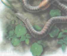
White planaria Habitat of the white planaria
In fresh water bodies you can find small, 1-2 cm long flatworms - planarias - hidden under snags, stones and leaves.
Bilateral symmetry of white planaria
The body of planarians is elongated and flattened from top to bottom. The rear end of the body is pointed, and the front is widened, and a short protrusion extends from it in both directions - these are the organs of touch, tentacles. Two black eyes are also placed here. If you look closely, you will notice that the right half of the planaria looks like a mirror image of the left. This symmetry, in contrast to the radial symmetry of coelenterates, is called bilateral. It is characteristic of most multicellular animals and arose in connection with the development of active movement.
Skin-muscle sac of white planaria
The body of the planaria is covered with cilia, thanks to which the planaria can move smoothly. Under the skin there are several layers of muscles. They do not lie in the form of separate bundles, but grow tightly together with the skin, forming skin-muscle sac. With the help of muscles, the planarian can change the shape of its body and move. There is no cavity under the skin-muscle sac, and the entire space between the organs is filled with loose connective tissue. Textile- is a union of homogeneous cells that perform a specific function. Thus, planarian muscle cells, similar in structure and function, make up muscle tissue that performs the function of movement. The tissue covering the animal’s body is called the integumentary tissue. Nerve cells combine to form nervous tissue. Thus, planarians have 4 types of tissues: cover, connecting, muscular And nervous. These tissues are present in all multicellular animals, more developed than flatworms.
Students make memorizing notes. Afterwards new groups are formed. Students talk in the group about the material they learned in the previous group.
After finishing the work, students create a mental map on the topic.
Students and the teacher fill out the table.
| Name of the planarian internal organ system | Organs forming the system | Functions of the organ system |
| 1.nervous | Head nerve ganglia and longitudinal nerve trunks connected by nerve bridges | Perception of irritations from the environment and from internal organs. The body's response to irritation |
| digestive | Mouth, pharynx, intestines | Capturing and digesting food |
| 3. excretory | Flame cells Excretory tubules, channels and pores. | Removal of liquid metabolic products |
| 4.sexual | Testes are tubular vas deferens. Paired ovaries-oviducts | Reproduction, increase in numbers |
Consolidation.
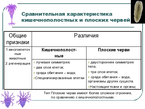
Students fill out the table in the notebook “Comparative characteristics of coelenterates and flatworms” and draw a conclusion from it.
They reveal features of a higher degree of organization of organisms in comparison with Coelenterates.
Homework.
Grading.
Formative assessment: Students applaud each other. Thank you for your work in class
Summative assessment: The teacher gives grades for the lesson.
Reflection.
Students fill out the table.
| I want to know |
||
- Equisetaceae department general characteristics and significance What structure does a horsetail spore have?
- Practical work “Structure of fern and horsetail. Horsetails have
- Who is behind the attacks on Tuleyev?
- Kirill Barabash - Lieutenant Colonel of the Air Force: biography, political views What is the IGPR “call”